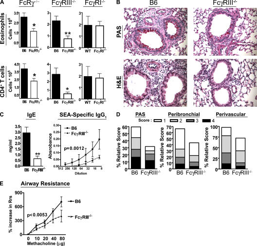
Figure 8.
FcγRIII−/− mice exhibit a decrease in Th2-mediated airway inflammation and airway hyperresponsiveness. (A) FcRγ−/− (n = 9), FcγRIII−/− (n = 8), FcγRI−/− (n = 8), and B6 mice were sensitized with i.p. injection of inactivated S. mansoni eggs on day 0. These mice were challenged with i.t. administration of SEA on day 7. On day 11, the mice were killed and the cellular composition of the BAL fluid was examined by flow cytometry (*, P < 0.05; **, P < 0.01). (B) Lung tissues from sensitized and challenged B6 and FcγRIII−/− mice were fixed in 4% formalin and embedded in paraffin. The sections were stained with PAS or hematoxylin and eosin as noted. (C) Amount of IgE and SEA-specific IgG1 in the sera of sensitized and challenged mice was estimated by ELISA (**, P < 0.01; P = 0.0012 obtained by two-way ANOVA analysis). (D) Goblet cell metaplasia, perivascular, and peribronchial inflammation were scored as described in Materials and methods. Four mice per group were analyzed. (E) Rrs to methacholine was measured at day 4 after challenge (P < 0.0053 by two-way ANOVA analysis). Data are representative of three independent experiments.

