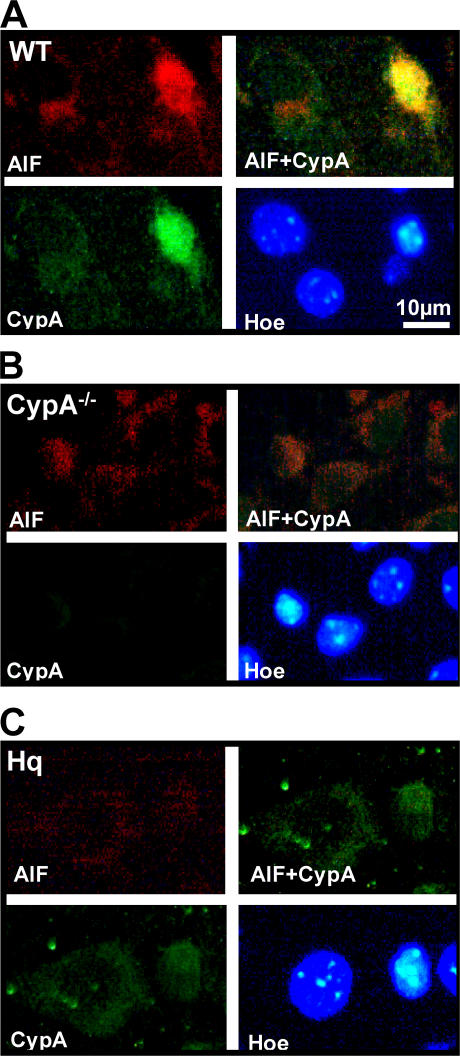
Figure 3.
Nuclear translocation of AIF was impaired in the absence of CypA and vice versa. (A) Immunofluorescent labeling of AIF (red), CypA (green), and chromatin (HOECHST 33342; blue) 3 h after HI in WT type mice showing both strong AIF and CypA nuclear staining in an injured neuron, as judged by the condensed nuclear morphology. (B) Labeling, as in A, in CypA−/− mice showing very weak nuclear AIF staining in injured neurons. (C) Labeling, as in A, in AIF-deficient Hq mice showing very weak CypA staining in injured neurons compared with WT type mice. Bar, 10 μm.

