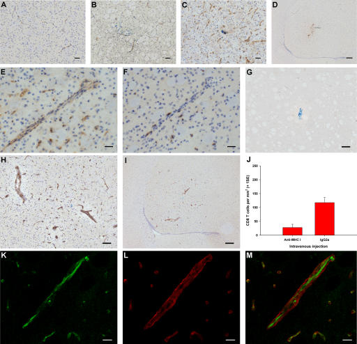
Figure 3.
The role of endothelial MHC class I in antigen-specific CD8 T cell infiltration into the brain. (A–F) Immunohistochemistry (brown) for MHC class I (A–E) and CD8 (F) in naive striatum (A) and striatum from CL4 mice injected with Cw3 (B) or HA (C–F). E and F show serial sections. (G–I) Immunohistochemistry (brown) for biotinylated IgG 3 d after intrastriatal HA injection (day 0) in CL4 mice receiving i.v. biotinylated anti–MHC class I antibody (H and I) or control biotinylated IgG (G) on day 2. (J) CL4 mice injected with HA intrastriatally received an i.v. bolus of blocking anti–MHC class I antibody or control IgG on day 2 and were perfused on day 3. There was a 76% reduction (95% CI = −139.5 to −40.0) in CD8 T cell infiltration (two-tailed Student's t test, P = 0.002). (K–M) High power confocal micrographs after double immunofluorescence on striatum for biotin (green in K) and γ1-laminin (red in L) (merged in M) 3 d after intrastriatal HA injection (day 0) in CL4 mice receiving i.v. biotinylated anti–MHC class I antibody on day 2. Bars: K–M, 10 μm; E and F, 30 μm; A–C, 50 μm; G and H, 100 μm; D and I, 200 μm.

