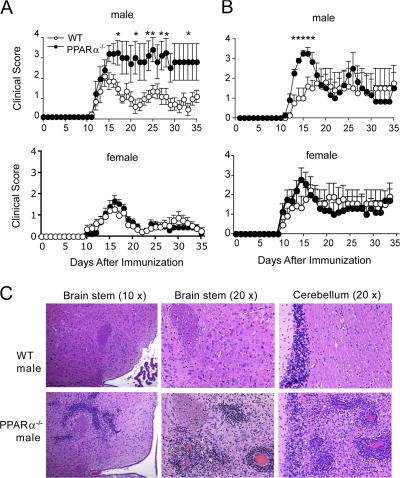
Figure 1.
Male PPARα−/− mice developed more severe clinical signs and displayed increased numbers of inflammatory brain lesions in the acute phase of EAE. Male and female WT or PPARα−/− SV.129 mice (n = 8–10/group) were immunized with MOG p35-55 in CFA and given intravenous injections of pertussis toxin on days 0 and 2 after immunization. (A and B) Mean + SEM clinical scores of mice in the different groups at various times after immunization. Shown are results of two independent EAE experiments. * indicates a significant difference from WT group (P < 0.05) as determined using a Mann-Whitney U statistic. (C) Paraffin-embedded sections of brain stem and cerebellum from representative male WT and PPARα−/− mice (from experiment no. 2) during the acute phase of EAE stained with hematoxylin and eosin. Bar in the bottom right, 50 μM.

