Figure 4.
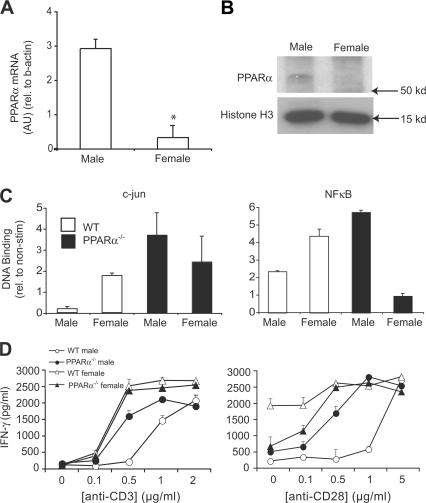
PPARα was more abundant in male as compared with female T cells and was associated with decreased NF-κB and c-jun activity and increased IFN-γ production. (A) Total RNA was obtained from naive CD4+ T cells that were pooled from the spleens of male and female SV.129 mice (n = 4 mice/group). The expression of PPARα mRNA in these cells was measured using real-time RT-PCR, and abundance was expressed relative to β-actin mRNA. Values are means ± SEM of PPARα/β-actin product abundance in triplicate reactions expressed in arbitrary units (AU). (B) Western blot analysis of PPARα in nuclear extracts (250 μg) prepared from male and female T cells. Histone H3 was used as a loading control. (C) c-jun (left) and NF-κB (right) DNA binding was measured in nuclear extracts from male and female SV.129 WT or PPARα−/− CD3+ T cells using an ELISA-based assay. Nuclear extracts were prepared from T cells at 16 h after stimulation with 5 μg/ml anti-CD3 and 5 μg/ml anti-CD28. Values are means ± SEM of absorbance units (duplicate culture wells) of stimulated wells expressed relative to absorbance in nonstimulated control wells. (D) CD3+ T cells from male and female SV.129 WT or PPARα−/− mice were stimulated with 0–2 μg/ml anti-CD3 (left) and 0–5 μg/ml anti-CD28 (right). IFN-γ production in culture supernatants was measured by ELISA at 72 h after stimulation. Values are means ± SEM of triplicate culture wells. Note that anti-CD28 and anti-CD3 were held constant at 0.5 μg/ml in the left and right panels, respectively. Results in A–D are representative of two independent experiments.

