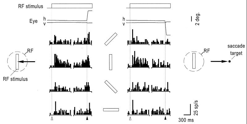
Figure 1.
Histograms showing the response of a V4 neuron after the appearance of an oriented bar in the RF and immediately before a saccadic eye movement either to the RF stimulus (Left) or a saccade target appearing 7° in the opposite hemifield (Right). In each column, spikes from the first half of the trial are aligned to the onset of the RF stimulus (▵) and then to the onset of the eye movement (▴) in 20 ms bins. Above each set of histograms are the mean horizontal (h) and vertical (v) eye traces. Bars between columns show the orientation of the RF stimulus. The RF of this neuron was located 4.2° from the fovea and centered on the horizontal meridian.

