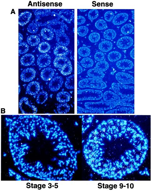
Figure 4.
In situ hybridization of mouse testis with PDE8. Approximate stage was determined by using Hoechst 33258 to stain nuclei. (A) Antisense and sense view under fluorescent light to observe the nuclear stain (blue) and bright field to observe probe signal (white dots). Note the low probe signal with the sense probe and the uneven probe signal within the semifierous tubules in the antisense field, indicating a spatial and temporal regulation of PDE8 expression. (B) Representative high magnification fluorescent and bright field of a early stage tubule and late stage tubule. Note the lack of probe signal (white dots) above background at stage 3–5 vs. the intense signal at stage 9–10.

