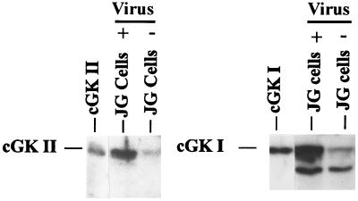
Figure 6.
Western blot of isolated mouse JG cells containing endogenous cGK II and cGK I (- virus), or additional cGK II and cGK I expressed from adenoviral vectors (+ virus). cGK II and cGK I were identified by their migration like recombinant kinases expressed in Sf9 cell lysates (86 and 76 kDa, respectively, shown at the left side of each panel) and were detected by using specific antisera and 125I- labeled protein A. The band migrating faster than cGK I is most likely a well known cGK I tryptic degradation product also sometimes observed in the cGK I standard. JG cell samples are 30 μg of protein each. The blots shown are representative of results from three different experiments.

