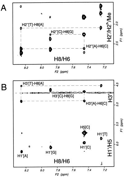
Figure 2.
Two-dimensional transferred NOESY spectra of an oligodeoxyribonucleotide. Two-dimensional transferred NOESY spectra of 0.80 mM d(TACG), 97 μM RecA protein, and 0.80 mM ATPγS at 180 msec mixing time at 25°C. The regions of H8/H6 and H2′/H2" are shown in A, and those of H8/H6 and H3′/H1′ in B.

