Figure 4.
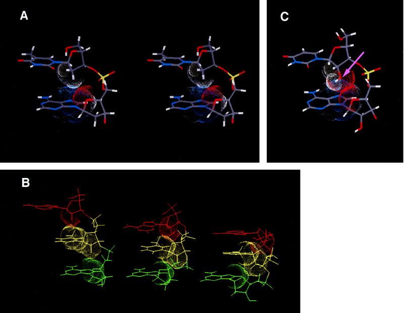
An extended single-stranded DNA structure induced by RecA protein. (A) Stereoview of the representative structure of RecA protein-bound DNA [sequence: d(TA)]. Van der Waals contact surface of a 2′-methylene moiety of a 5′ side residue (T) and those of O4′, C4, C5, N7, C8, H8, and N9 of the 3′ side residue (A) are shown. A structure deduced from the calculation by use of a simulated annealing method was refined using revised parameters as published (27). (B) Side views of the structure of DNA in RecA protein-bound form (Left), B-form (Middle), and A-form (Right). Van der Waals contact surfaces between adjacent residues are shown. In B- or A-form DNA the whole structure must be disrupted on the process of strand exchange because of its close packing between adjacent residues. (C) A hypothetical RNA structure in the RecA protein-bound form. H2" is replaced upon a hydroxyl group (indicated by arrow) in RNA.

