
Fig. 4.
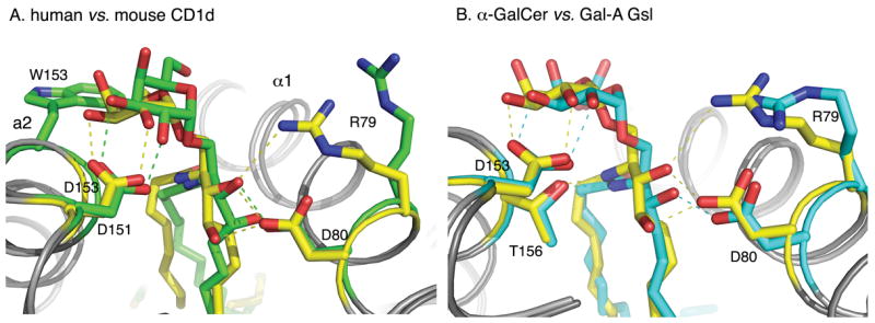
CD1d hydrogen-bonding network. The CD1d ligands (yellow, green and cyan) are stabilized by hydrogen-bonds (dashed lines) between the α1 and α2-helices of either human CD1d (A) or mouse CD1d (B). (A) Comparison of α-GalCer presentation by mouse (yellow) and human (green) CD1d. The ligands are in the same color as the CD1d molecules they are bound to, in order to emphasize the overall conformational change in the ligand-CD1d complexes. The most dramatic difference is W153 in human (G155 in mouse), which tilts and pushes the galactose towards the center of the CD1d binding grove. (B) Comparison of α-GalCer (yellow) and GalA-Gsl (cyan) binding to mouse CD1d. GalA-Gsl is inserted deeper into the F′ pocket and, as a result, the galacturonic headgroup is laterally shifted 1 Å towards the center of the binding groove.