
1.
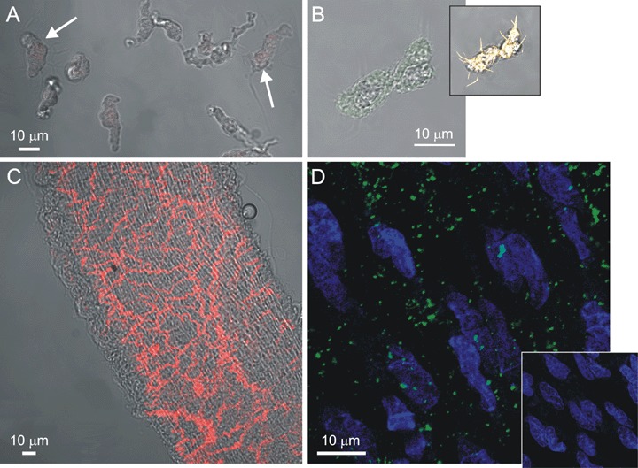
Fluorescent staining for PGP9.5 and vWF. (A) A combined image of transmitted light (shades of grey) and PGP9.5 fluorescence (red) of VSMCs and AIL cells (arrows) obtained by confocal microscopy. Neither AIL cells nor VSMCs stained positive for PGP9.5 (n = 24 cells). (B) A combined image of transmitted light (shades of grey) and vWF fluorescence (green) of an AIL cell, showing only weak non-specific staining (n = 6). Inset: Same cell stained with BODIPY 558/568 phalloidin (yellow) revealed the presence of otherwise faintly visible filopodia, confirming the identity of an AIL cell. (C) Same as in (A), but a segment of mesenteric artery was used, showing the presence of nerve fibers (positive control, n = 5). (D) A fluorescent image of an endothelial layer in mesenteric artery segment immunostained for vWF and stained with SYTO 40 for nucleic acids. Nuclei of endothelial cells can be seen in blue. Green colour shows vWF staining of granular appearance, due to its storage in the Weibel-Palade bodies of endothelial cells (n = 4). Inset: As for the main panel, but the primary antibodies to vWF were omitted.