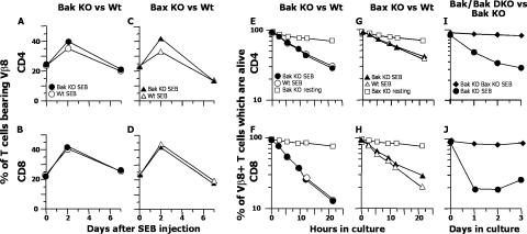
Figure 1.
Activated T cell death requires either Bak or Bax. (A–D) BL6, Bak KO, Bax KO, and Bax+/+ littermates of Bax KO mice were injected with 100 ug SEB i.v. At the indicated times, cells were harvested from spleens and the percentage of CD4+ or CD8+ T cells bearing Vβ8x was determined. Closed and open symbols represent the results for KO and wild-type mice, respectively. Results shown are means ± standard deviations of data from three mice at each time point. (E–J) BL6, Bak KO, Bax KO, Bax+/+, and RAG KO mice reconstituted with Bak KO or Bak KO Bax KO bone marrow were given SEB. 63 h later, T cells were purified from their spleens and lymph nodes and cultured. At various times thereafter, the percentages of CD4+ and CD8+ Vβ8+ T cells that were alive, as defined by their light scatter properties (reference 29), were determined. (E and F) •, SEB-injected Bak KO; ◯, SEB-injected BL/6; □, uninjected Bak KO cells. (G and H) ▴, SEB-injected Bax KO; ▵, SEB-injected Bax+/+ littermates; □, uninjected Bax KO cells. (I and J) ♦, SEB-injected Rag KO mice reconstituted with Bak KO Bax KO fetal liver cells; •, SEB-injected Rag KO mice reconstituted with Bak KO fetal liver cells. (E, G, and I) Results for CD4+ T cells. (F, H, and J) Results for CD8+ T cells. Results shown are means ± standard deviations of data from three samples analyzed at each time point in one experiment. Results are also illustrative of two independent experiments for the Bak KO Bax KO T cells and of more than four independent experiments for the Bak KO and Bax KO cells.

