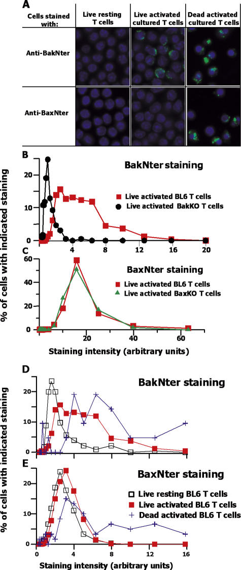
Figure 2.
Before activated T cells die, a considerable amount of Bak, but not Bax, changes its shape. BL6 mice were injected with 100 ug SEB. 2 d later, T cells were isolated from their spleens and lymph nodes and cultured for 7 h. The cells were then sorted based on PI staining to obtain Vβ8+ live and dead populations, and stained for NterBak or NterBax. (A) Typical fields of the different types of cells are shown. (B–E) Individual cells were ringed and analyzed for their extent of NterBak (B and D) or NterBax (C and E) staining and surface area. The level of staining of each cell was corrected for background levels of staining, with the following calculation: [(the amount of staining with the particular antibody/surface area) − (background staining/surface area)] × surface area. The cells were then assigned to bins based on their level of staining with each antibody. Results shown are as follows: (the number of cells in each bin/total number of cells analyzed) × 100. Results represent analyses of at least 226 live cells and 21 dead cells of each type. The experiments shown in B and D and in C and E were performed at different times. Settings and conditions were slightly different between the two experiments shown, which led to differences in the level of anti-Bax staining of activated cells.

