Figure 1.
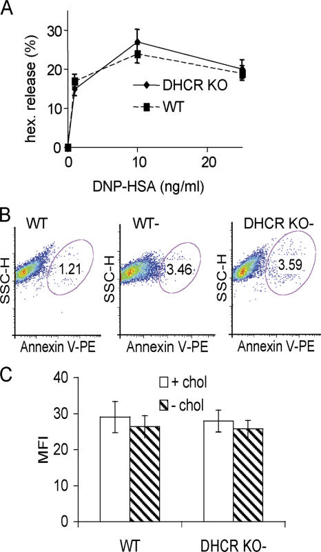
Characterization of mast cell FcɛRI expression, survival, and function from DHCR KO mice. (A) Mast cells derived from the liver progenitors of WT or DHCR KO mice were cultivated in medium with normal FBS, IL-3, and stem cell factor for 4 wk. Cells were sensitized with DNP-specific IgE for 60 min and activated by exposure to the indicated concentration of DNP-HSA for 20 min. Hexosaminidase release is expressed as a percentage of total cellular hexosaminidase. Data is mean ± SEM from four individual experiments. (B) Apoptotic cells were stained with PE-labeled annexin V in WT nondepleted or WT and DHCR KO cells depleted in cholesterol-free medium for 72 h and analyzed by FACS. One representative of >12 individual cultures is shown. (C) WT or DHCR KO mast cells were cultivated in normal or cholesterol-free medium for 72 h and loaded with IgE, washed, and followed by FITC-labeled anti-IgE antibody. Level of FcɛRI expression was determined as the mean fluorescence intensity (MFI) in FITC anti-IgE–stained cells compared with isotype control. Data is mean ± SD from three individual experiments.

