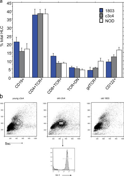
Figure 4.
Cellular composition of the HLC population changes with age in NOD.c3c4, but not strain 1803, mice. (a) The NOD.c3c4 HLC population does not differ from that of NOD or 1803 mice before 30 wk of age. HLCs were isolated, stained with surface markers, lymphocyte gated, and analyzed by FACS (see Materials and methods) from NOD, 1803, and NOD.c3c4 mice. There was no significant difference in cell composition between NOD, NOD.c3c4, or 1803 mice (1803 NOD, n = 3–5; NOD.c3c4, n = 8–10, except in the case of γδT; NOD.c3c4, n = 4). All mice were 15–25 wk old. (b) Granulocyte accumulation in aged NOD.c3c4 mice. In NOD.c3c4 and 1803 >30 wk old, HLCs were isolated and analyzed by FACS as described above, using granulocyte gates (circled areas). Aged NOD.c3c4 showed a significantly increased accumulation of GR-1+ cells (41.4%) compared with aged 1803 mice (7.8%), which did not differ from young NOD.c3c4 mice (7.4%).

