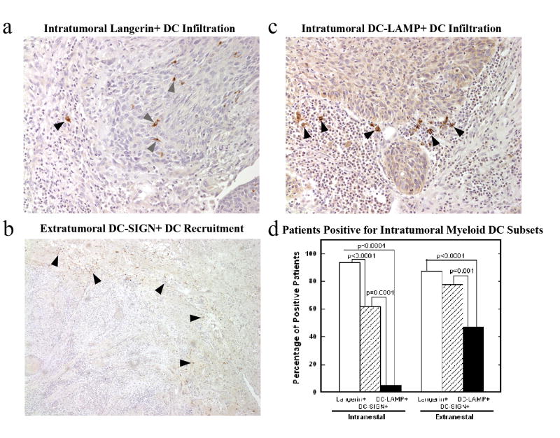
Figure 1. Myeloid dendritic cell subsets display different patterns of primary tumor infiltration.
Immature Langerin+ dendritic cells were predominantly present within the tumor area, both intranestal (
 ) and extranestal (▲), 20x (a). Immature DC-SIGN+ dendritic cells were predominantly present outside the tumor area (▲), 5x (b). Mature DC-LAMP+ dendritic cells (▲) 20x (c), were present in fewer patients than the immature DC subsets (d).

