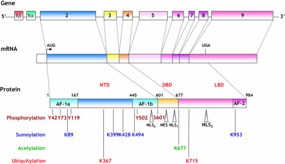
Figure 1. Schematic representation of human MR structure.
MR gene, mRNA, protein, functional domains and associated posttranslational modifications are depicted. The hMR gene is composed of ten exons, including two untranslated first exons (1α and 1β). The AUG translational initiation start codon is located 2 bp after the beginning of exon 2, while the stop codon is located in exon 9. Multiple mRNA isoforms generated by alternative transcription or splicing events are translated into various protein variants, including those generated by utilization of alternative translation initiation sites (not shown). The receptor is comprised of distinct functional domains (activation function AF-1a, AF-1b and AF-2) and nuclear localization signals (NLS0, NLS1 and NSL2), as well as one nuclear export signal (NES). The positioning of amino acids targeted for phosphorylation, sumoylation, acetylation and ubiquitylation is indicated for the human MR sequence.

