
Figure 6. A hierarchical docking result showing a low energy configuration of PNA (magenta) and 2EN (green).
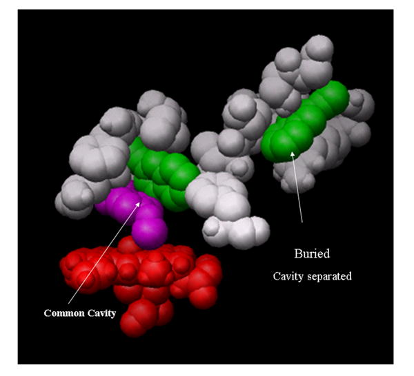
A single 2EN and the PNA are in a “common-cavity” arrangement above the heme (red) shown in the common cavity. A second 2EN is shown in a “separate cavity” adjoining the heme binding site. The residues within 2.8 Å of 2EN and PNA in the closed cavity arrangement are PNA [E301,V367,I363,V477,G478] and 2EN[I101,V104, I114,I209,F297,E301,V367,V477]. The residues within 2.8 Å of 2EN and PNA in the separated cavity configuration are: PNA[V104,F115,F297,E301,I363,V367] and 2EN[H172,F203,F206,S207,S210, T306,L362,G478,N479,V480,I486]. Residues have been removed for clear visualization of the occupancy; both of these sites are buried.