
Figure 4.
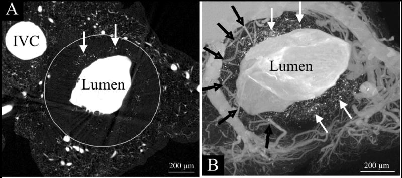
Micro-CT transaxial images of a double knockout mouse (LDL-/-, apoE-/-) aorta after it was injected with a radiopaque silicon polymer. The aortic lumen (large white area) is irregular rather than round because of the atherosclerotic plaques encroaching into the lumen. The small vessels around the lumen are vasa vasorum. Note that unlike the “clear” zone between the vasa vasorum and the main lumen in the normal artery (upper panels of Fig. 1), the black arrows in this image show vasa vasorum entering the plaque area close to the main lumen surface. These are the newly formed vasa vasorum in response to the plaque formation process. Also, the white arrows point to isolated punctate opacities in the region of the plaques. These were shown to be small accumulations of iron and calcium, presumably the remains of red blood cells. [Abridged version of Figure 3 with permission from Langheinrich AC et al., Ref. 109].