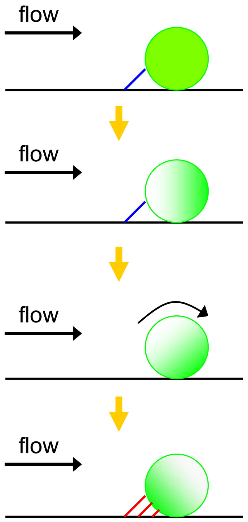
Figure 4. Proposed shear-induced L-selectin cap formation model for E-selectin mediated neutrophil rolling under flow.
The E-selectin tether with PSGL-1 is represented as a blue line and the E-selectin tether with L-selectin is represented as a red line. The green shades indicate L-selectin distribution on a cell and the yellow arrows indicate possible sequences of events. In the top diagram, a long-lived E-selectin:PSGL-1 tether results in a cell pause. During the cell pause, shear applied on the cell membrane induces L-selectin cap formation. As the applied shear force continues, the E-selectin:PSGL-1 tether dissociates, followed by rotation of the cell. Rotation allows new multiple adhesive interactions of E-selectin with the concentrated L-selectin cap to firmly arrest the cell. Eventually, localized L-selectin is observed upstream in neutrophil rolling experiments on E-selectin (bottom row).

