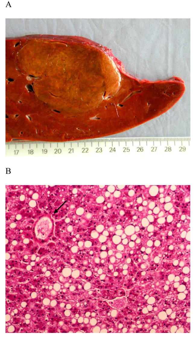
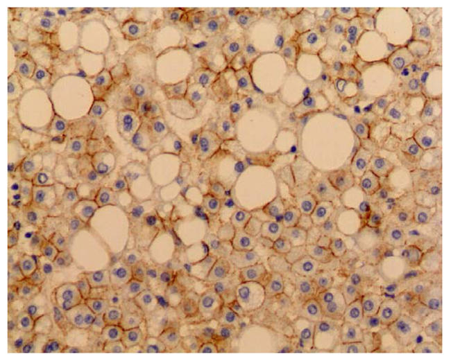
Figure 1.


A: Macroscopy: the liver lesion measured 7 cm, was soft, yellow, well demarcated with no hemorraghe or necrosis, B: Microscopy: the lesion was composed of benign appearing hepatocytes with marked steatosis, intermingled with thin walled vessels (arrow) (HES staining, original magnification ×100) C: β-catenin immunostaining: hepatocytes plasma membrane staining with no nuclear reactivity in the hepatocellular adenoma (original magnification ×200)
