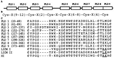
Figure 3.
Domain structure of the pacifastin light chain. (A) Schematic diagram showing the positions of the nine PLDs. (B) The common cysteine spacing pattern of the nine PLDs, and LICM I, LICM II (11), HI (14). (C) Alignment of the nine PLDs, LICM I, LICM II, and HI. Conserved cysteines are in boldface. Underlined residues indicate putative reactive site, P1-P′1 of the three locust inhibitors (14). Gaps (−) are introduced to optimize the alignment.

