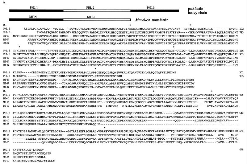
Figure 4.
Structure of the pacifastin heavy chain. (A) Schematic diagram showing the three PHLs in comparison with Manduca transferrin (8). Filled boxes represent sequence similarity to the N-terminal lobe of transferrins and open boxes represent similarity to C-terminal lobe of transferrins. (B) Alignment of PHL 1 and PHL 3 with N-terminal lobe of transferrins from B. discoidalis, BT-N (9), M. sexta, MT-N (8), and S. peregrina, ST-N (10). (C) Alignment of PHL 2 with C-terminal lobe of transferrins from B. discoidalis (BT-C), M. sexta (MT-C), and S. peregrina (ST-C). Conserved residues that are essential for iron-binding are in bold and indicated with an ∗. Conserved cysteines are in bold. Gaps (−) are introduced to optimize the alignment.

