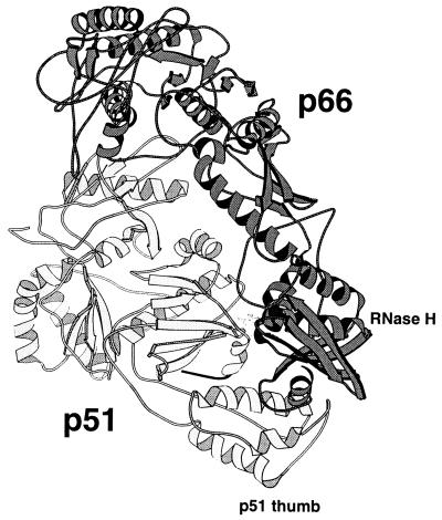
Figure 1.
Structure of HIV RT. The p66 subunit of unliganded RT is darkly shaded, and the p51 subunit is light except for the carboxyl-terminal residues (426–434) drawn in black (9). Six residues (435–440) at the extreme carboxyl terminus of p51 were not present in the crystal structure and are not shown. The RNase H domain of p66 and the thumb domain of p51 are indicated. The drawing was produced with the program molscript.

