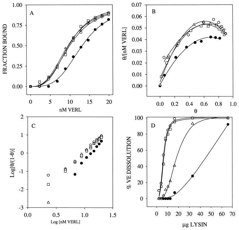
Figure 3.
Lysin–VERL binding is consistent with lysin-mediated VE dissolution. (A) Fluorescence polarization of VERL–lysin binding. The sigmoidal binding curves indicate positive cooperativity, as supported by convex Scatchard plots (B) and Hill plots with slopes of approximately 3 (C). (B and C) θ = the fraction of lysin bound. (D) Lysin-mediated VE dissolution measured by light scattering. Species combinations are: □, Hr VERL + Hr lysin; ○, Hc VERL + Hc lysin; ▵, Hr VERL + Hc lysin; •, Hc VERL + Hr lysin. The species selectivity in lysin–VERL binding curves (A) is consistent with the species selectivity of lysin in dissolving isolated VEs (D).

