
Figure 2.
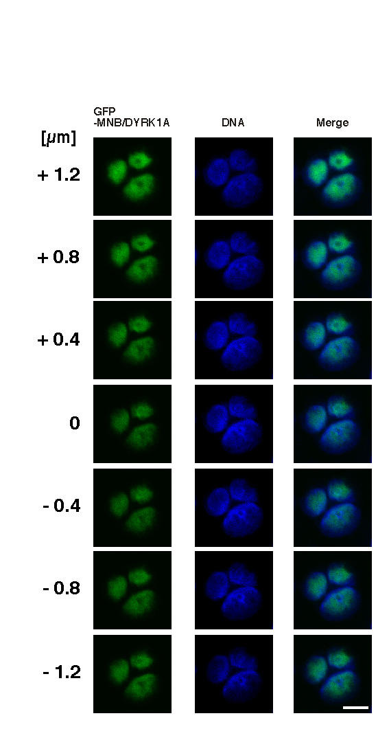
Appearance of multiple nuclei in GFP-MNB/DYRK1A-overexpressing cells. HeLa cells transfected with the plasmid encoding GFP-MNB/DYRK1A were incubated for 48 hours and then fixed as described in "Materials and Methods." Fixed cells were stained with 1 μM TOTO-3 for the counterstaining of DNA and observed by confocal laser scanning microscopy. Images were collected at 0.4-μm Z axis intervals. GFP-MNB/DYRK1A (left) and DNA (middle) are displayed in green and blue, respectively. Merged images are shown in the right panel. Scale bar, 10 μm.