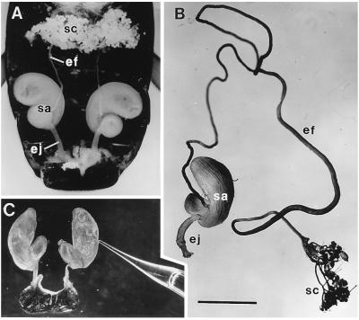
Figure 1.
(A) Dorsal view of abdominal cavity of G. janus, showing the two defensive glands. Other abdominal organs have been largely excised. Each gland consists of an aggregate of secretory cells (sc), an efferent duct (ef), a storage sac (sa), and an ejaculatory duct (ej). (B) Isolated gland of G. lecontei (labels as in A). (C) Excised glandular sacs of G. lecontei, being “milked” of secretion; one of the sacs has been pierced with a glass micropipette, which is taking up secretion by capillary action. (Bar: 2 mm.)

