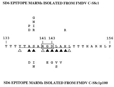
Figure 1.
Alignment of the amino acid sequences of the central region of the VP1 G–H loop of FMDV C-S8c1 and SD6 epitope MARMs. The single-letter amino acid code is used. Numbering is according to that for FMDV C-S8c1. Antigenic site A of type C viruses involves residues 136–150. The continuous epitope recognized by mAb SD6 is underlined. The RGD triplet is boxed. Solid triangles correspond to amino acid residues that cannot be replaced by most other amino acids with regard to binding to SD6; open triangles indicate residues that can be replaced by some other residues without having a drastic effect on SD6 recognition; residues 137 and 140 were generally replaceable without affecting SD6 recognition (ref. 33 and M.G.M. and E.D., unpublished work). These results are also consistent with structural data regarding the mAb SD6–G–H loop peptide interaction (23).

