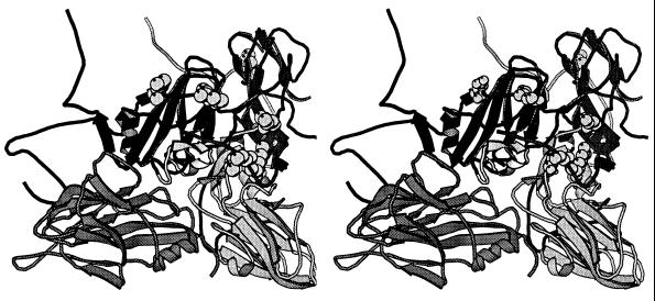
Figure 2.
Location of the amino acid substitutions found in FMDV C-S8c1p100c10 on the virus capsid. Stereoview of a ribbon protein diagram of a crystallographic protomer of C-S8c1 in which the relevant substitutions have been modeled (see Materials and Methods). The capsid proteins VP1, VP2, and VP3 are indicated as dark, medium, and light grey, respectively. VP4 is entirely internal and has not been included for clarity. VP1 from a neighboring protomer is shown on the left side (dark grey). The substituted residues are depicted in CPK representation. The position of the G–H loop of VP1 in C-S8c1 complexed with mAb SD6 is indicated in light grey (37).

