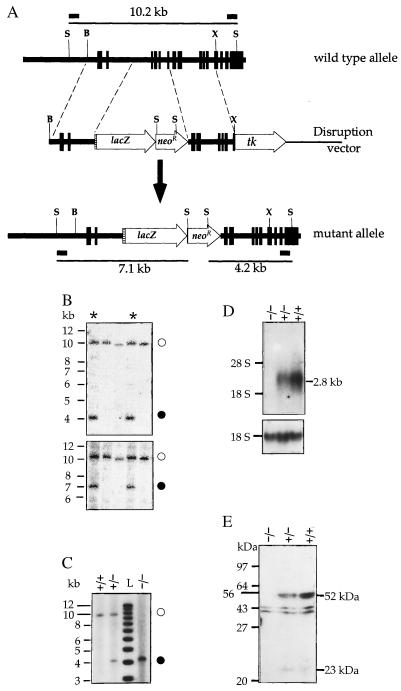
Figure 1.
(A) Disruption of the Pcsk4 gene. Vertical boxes represent exons, the hatched area an acceptor splice site, and short horizontal bars DNA probe locations. B, BstZI; X, XhoI; S, SphI. (B and C) Southern blot genotyping of ES cell clones (B) and mouse tail tips (C) with a 3′ (B Upper and C) or the 5′ probe (B Lower). Open circles, solid circles, and asterisks identify the wild-type allele, the mutant allele, and recombinant clones, respectively. L stands for 1-kb ladder. (D) Northern blot analysis of testicular RNA of wild-type (+/+), heterozygous mutant (−/+), and homozygous mutant (−/−) mice for PC4 mRNA (Upper) and 18S rRNA (Lower). (E) Western blot analysis of epididymal sperm. The two immunoreactive proteins of about 52 and 23 kDa detectable in wild-type and heterozygote mouse samples are PC4 related. They were not observed after the antiserum had been preabsorbed with purified PC4 (not shown), unlike the two proteins of about 40 and 44 kDa detectable in all three samples, which are likely due to a nonspecific immunoreaction.

