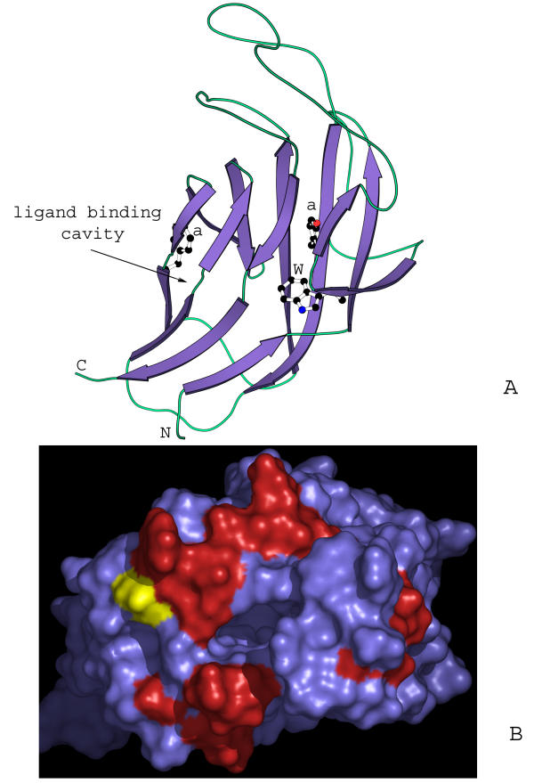
Figure 3.
Models of 7TMR-DISMED1 and the TM domain of the 7TMR-DISMs. (A) Prototype of the β-jellyroll seen in 7TMR-DISMED1, sialate 9-O-acetylesterases, β-glucoronidases and β-glucosidases. The β-jellyroll domain shown here is a cartoon representation of the domain from the crystal structure of β-galactosidase (PDB:1GHO). Conserved residues typical of the 7TMR-DISMED1 are shown in ball stick representation. "a" stands for a conserved aromatic position. (B) A homology model of the TM domain of the 7TMR-DISMs showing the distribution of conserved residues in the 7TMR-DISMs with 7TMR-DISMED1 domains. The model was constructed using bacteriorhodopsin (PDB: 1C3W) and bovine retinal rhodopsin (PDB:1F88) as templates. The N terminus of the 7TMR-DISM domain, where the extracellular domain is attached, is shown in yellow. The red color shows the distribution of residues on the external surface, which are uniquely conserved in 7TMR-DISMs with 7TMR-DISMED1s. This set of proteins essentially corresponds to the lower group of sequences in Fig. 1.

