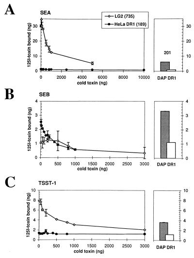
Figure 1.
SEA, SEB, and TSST-1 binding to HeLa DR1 and LG2. Specific and saturable binding is demonstrated when increasing amounts of cold toxin successfully displace bound 125I-labeled SEA (A), SEB (B), or TSST-1 (C) as described (7). HLA-DR expression was analyzed by cytofluorometry using the anti-DR antibody L-243. The numbers indicate the mean of fluorescence for HLA-DR expression on all three cell types. (Right) The hatched bars represent the binding of the same amount (100 ng) of SEA (A), SEB (B), or TSST-1 (C) on DAP DR1 cells as a control. Open bars represent the binding of 100 ng of toxin in the presence of a 10-fold excess (1 μg) of cold toxin. Standard variations for each point (duplicates) is represented by the error bars. Because we do not detect binding of SEB on LG2, we approximate the dissociation constant (Kd) of the interaction to be ≥10−5 M.

