Figure 3.
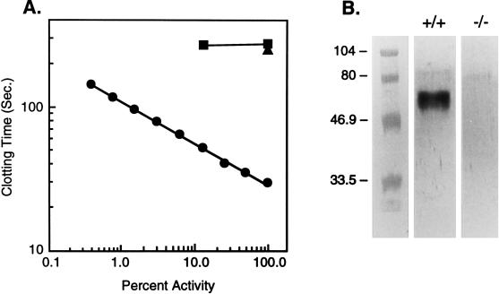
Verification of TF gene disruption. (A) TF activity in TF(+/+) (•) or TF(−/−) (▪) immortalized MEFs. Data are from a single representative experiment. Cell lysates were diluted serially in TBSA from 1:4–1:512, and TF activity was assessed in a one-stage clotting assay as described (28). The negative control has the cell lysate replaced with TBSA (50 mM Tris, pH 7.5/100 mM NaCl/0.1 mg/ml BSA) (▴). Data are plotted as percentage of activity vs. clotting time in seconds. The activity in a TF(+/+) cell lysate diluted 1:4 is designated as 100% activity. (B) Western blot analysis, using an anti-mouse soluble TF polyclonal antibody, of immortalized MEF cell lysates. Lane (+/+) is of a wild-type cell lysate, showing mTF migrating at ≈50 kDa, which is comparable to published data (12). Lane (−/−) is of a TF-deficient cell lysate, showing neither TF nor an aberrant protein product.

