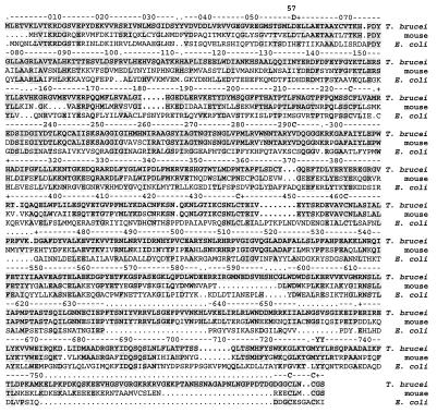
Figure 2.
Alignment of the T. brucei R1 protein with the mouse (48) and E. coli (45) class Ia R1 proteins. The ruler follows the conventionally used numbering of the E. coli R1 protein (45). Breaks in the alignments are indicated with dots; breaks in the E. coli R1 sequence are indicated as dots in the ruler as well. Unless written out, every tenth residue is indicated as a cross in the ruler. Shaded residues are identical to the T. brucei R1 protein amino acid sequence. Residues mentioned in the text are indicated as the one-letter abbreviation. These are the active site cysteines (C-225, C-439, C-462), the residues in the electron transport pathway (Y-730, Y731), the cysteines that shuttle electrons from thioredoxin or glutaredoxin (C-754, C-759), and the allosteric effector binding-site residues (D-57, C-292). Mouse R1 protein amino acid numbering is used for the allosteric activity site residue (D-57). Otherwise, E. coli R1 protein amino acid numbering is used.

