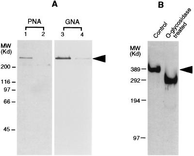
Figure 3.
Identification of carbohydrate structures on the IIM by lectin binding assays and glycosidase treatments. (A) Lectin binding analysis. IIM was electrophoresed by SDS/PAGE, transferred onto blotting membrane and assessed for terminal mannose and galactose β(1–3) N-acetylgalactosamine with G. nivali agglutinin (GNA) (lane 3) and peanut agglutinin (PNA) (lane 1), respectively. Lane 2 shows binding of PNA to IIM pretreated with O-glycosidase. Lane 4 shows binding of GNA to IIM pretreated with N-glycosidase F. (B) SDS/PAGE analysis of the IIM treated with O-glycosidase, showing an apparent molecular weight reduction from 400 to 300 kDa (left lane, control; right lane, treated) (silver-stained gel). The position of IIM in both A and B are indicated by an arrow.

