
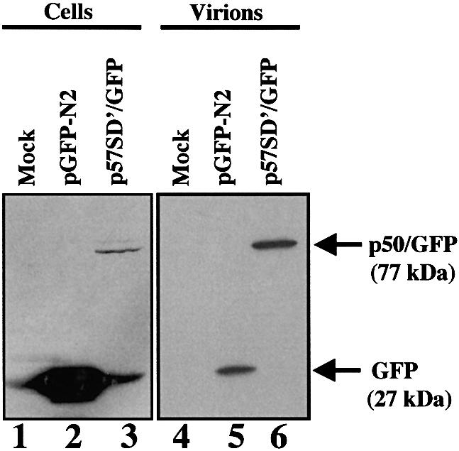
Fig. 8. Detection of p50/GFP protein in MLV particles. Same amount of NIH3T3 infected cells were transfected with p57SD′/GFP or pGFP-N2 vector. To allow quantitative analysis proteins were extracted from 5 × 10⁵ cells (lanes 1–3) and from corresponding cell-free pelleted supernatant (lanes 4–6) and analyzed by western blotting using an anti-GFP antibody. Mock-transfected cells (lanes 1 and 4) and cells transfected with GFP vector (lanes 2 and 5) were used as controls.