
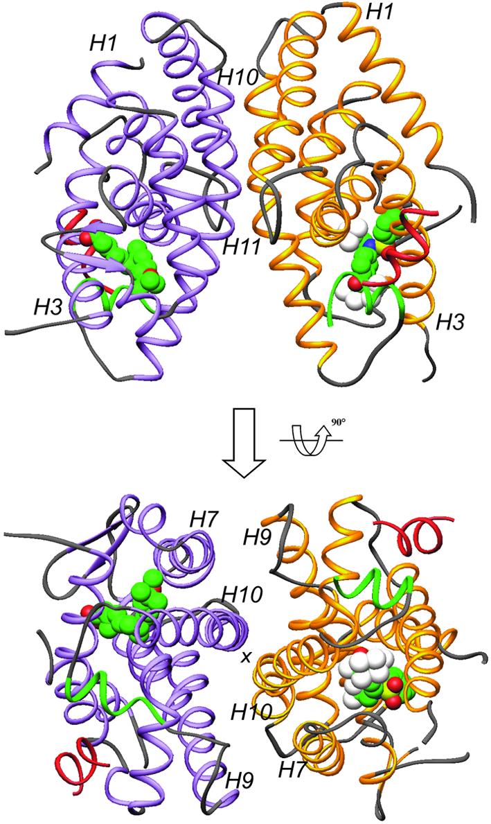
Fig. 1. Overall structure of the LXRα–RXRβ LBD-heterodimer presented in two views separated by 90°. The LXRα LBD is shown in yellow and the RXRβ LBD in purple, except for AF2 helices which are depicted in green. GRIP-1 peptides are colored red. T-17 and MPA are in space-filling representation, with carbon, oxygen, nitrogen, sulfur and fluoride atoms colored in green, red, blue, yellow and white, respectively. Selected secondary elements are annotated with numbers positioned at their N-terminal ends.