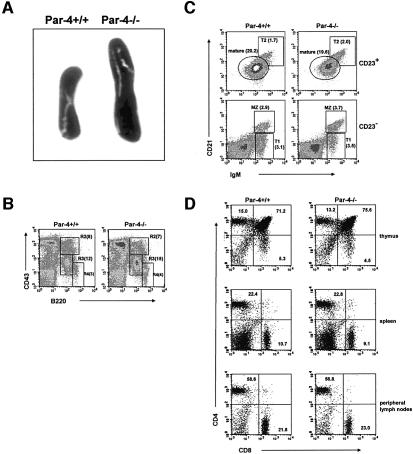
Fig. 1. Immunological phenotypic analysis of Par-4-deficient mice. (A) Representative spleens (of >20 mice) from WT and Par-4–/– mice. (B) Single cell suspensions of bone marrow cells from WT and Par-4–/– mice were stained with anti-CD43 versus anti-B220. R2, pro-B; R3, pro-B and pre-B; R4, mature B cells. Values in parentheses correspond to the percentage of each subpopulation. (C) Three-color flow cytometric analysis of splenocytes from WT and Par-4-deficient mice were carried out by staining with antibodies to IgM, CD21 and CD23. Cells were separated into CD23-positive and -negative; MZ (CD21hiIgMhiCD23–), marginal zone B cells; mature B cells are characterized by CD21intIgMlowCD23+; T1 (CD21lowIgMhiCD23–), transitional 1 immature B cells; T2 (CD21hiIgMhiCD23+), transitional 2 immature B cells. Values in parentheses correspond to the relative percentage of each subpopulation. (D) Single cell suspensions of thymuses, spleen and peripheral lymph nodes from WT and Par-4-deficient mice were stained with anti-CD4 versus anti-CD8. Results in (B–D) are representative of four experiments with similar results.

An official website of the United States government
Here's how you know
Official websites use .gov
A
.gov website belongs to an official
government organization in the United States.
Secure .gov websites use HTTPS
A lock (
) or https:// means you've safely
connected to the .gov website. Share sensitive
information only on official, secure websites.
