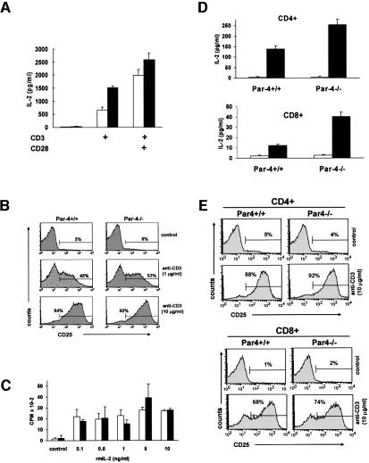
Fig. 3. IL-2 secretion and proliferation in Par-4-deficient T cells. (A) IL-2 production in supernatants of T cells from WT and Par-4–/– mice activated (black bars) or not (empty bars) with 10 µg/ml of anti-CD3. (B) Surface expression of CD25 was analyzed by flow cytometry 24 h after stimulation of WT or Par-4 KO T cells with different concentrations of anti-CD3. (C) T cells from WT (empty bars) or Par-4 KO (black bars) mice were stimulated with 0.1 µg/ml of anti-CD3 antibody in the absence or in the presence of increasing concentrations of recombinant murine IL-2 (rmIL2) for 72 h. (D) In another set of experiments, IL-2 production by non-activated (empty bars) or activated (10 µg/ml anti-CD3; black bars) purified CD4+ (upper panel) or CD8+ (lower panel) T cells from WT or Par-4–/– mice was determined as described above for total T cells. (E) CD25 surface expression of non-activated or activated (10 µg/ml anti-CD3) purified CD4+ (upper panel) or CD8+ (lower panel) T cells from WT or Par-4–/– mice was determined as described above for total T cells. The experiments in (B) and (E) are representative of another two with similar results and those in (A), (C) and (D) are the mean ± SD of three independent experiments with incubations carried out in duplicate.

An official website of the United States government
Here's how you know
Official websites use .gov
A
.gov website belongs to an official
government organization in the United States.
Secure .gov websites use HTTPS
A lock (
) or https:// means you've safely
connected to the .gov website. Share sensitive
information only on official, secure websites.
