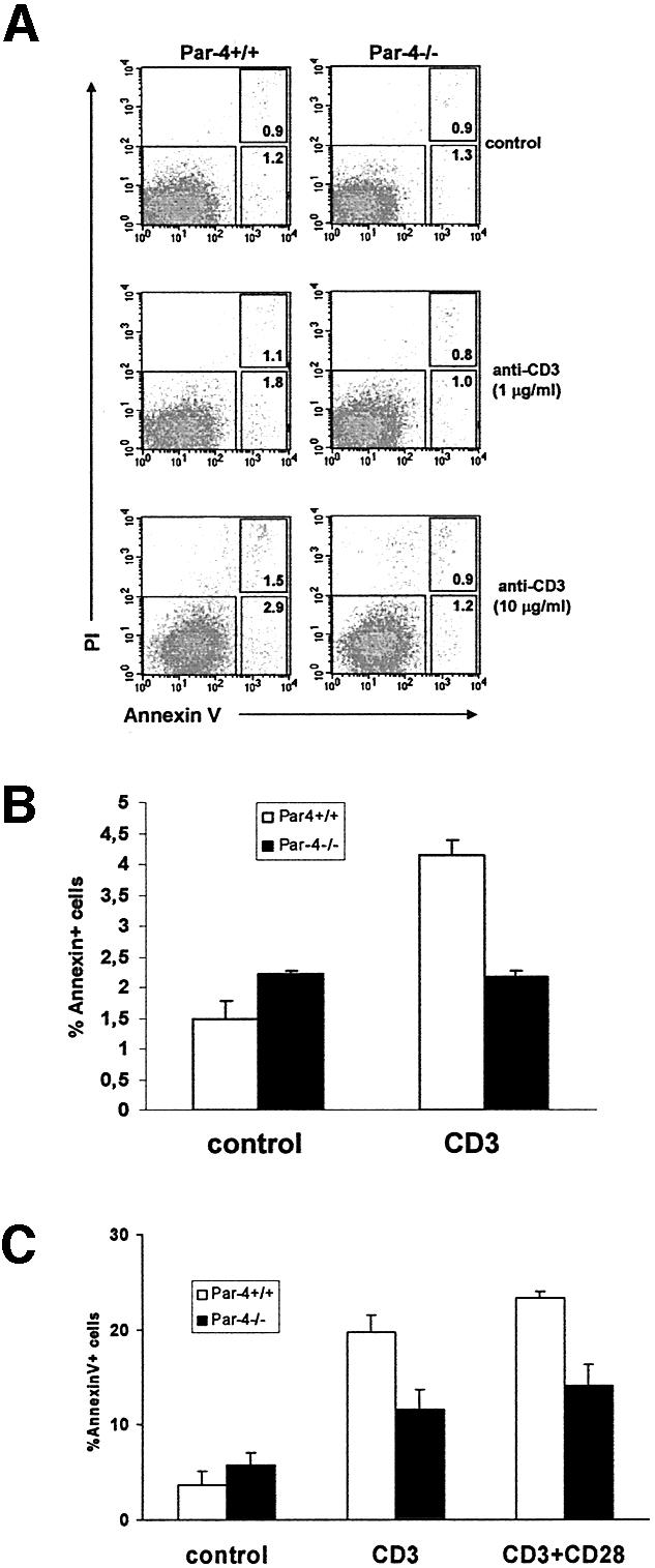
Fig. 4. T-cell apoptosis. (A) T cells from WT and Par-4 KO mice were stimulated with different concentrations of anti-CD3 antibody for 24 h, stained for the binding of annexin V and analyzed by flow cytometry gated for viable cells; the right upper inset corresponds to cells undergoing early apoptosis whereas the right lower inset corresponds to cells at a more advanced stage of the apoptotic process. Shown is a representative experiment of another three experiments with incubations carried out in duplicate. (B) The mean ± SD of the total (early + late) apoptosis of these experiments. (C) In another set of experiments, T cells, either WT or Par-4-deficient, were stimulated with anti-CD3 (3 µg/ml) in the presence of rIL-2 for 48 h, after which activated T cells were harvested, washed and re-plated, followed by the addition of anti-CD3 or anti-CD3 plus anti-CD28. Cell viability was determined as above at 2 days post-stimulation.
