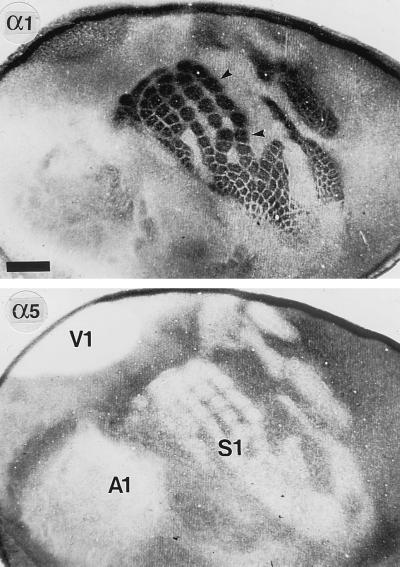
Figure 1.
Complementary distribution of the α1 and α5 subunits in the neocortex at P7, as seen in adjacent tangential sections through layers III–IV processed for immunoperoxidase staining with subunit-specific antibodies. To prepare these sections, the cortex was dissected free of subcortical tissue, flattened between two glass coverslips, and postfixed for 48 hr at 4°C. Sections were then cut parallel to the pial surface and processed for immunoperoxidase staining (intense signals appear black). The localization of the three major primary sensory areas S1, V1, and A1 (the primary auditory area) is revealed by the complete lack of α5 subunit staining in layer IV. Intervening association areas are moderately stained for the α5 subunit. By contrast, the α1 subunit IR is particularly prominent in S1 and V1 and reveals in great detail the somatotopic organization of the barrel field (arrowheads). In the remaining neocortex, including A1, the α1 subunit is weak to moderate. The reciprocal expression pattern of the α1 and α5 subunits is best seen in S1, where patches of intense α1 subunit staining are matched by corresponding patches devoid of α5 subunit IR. (Scale bar = 1 mm.)

