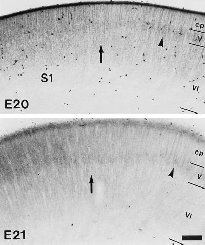
Figure 4.
Area-specific distribution of the α1 subunit IR in fetal neocortex. The photomicrographs illustrate the lateral boundary of S1 (arrows) as seen in transverse sections at E20 and E21 in sections processed for immunoperoxidase staining. In the territory corresponding to the future area S1, the α1 subunit IR is initially diffuse and uniform across the cortical plate (cp), whereas in the adjacent cortex, it is confined to the developing layer V (arrowhead), with a narrow transition zone in between. These areal boundaries are more prominent at E21, because the deeper cortical layers appear more differentiated and the α1 subunit IR increases in S1. (Scale bar = 100 μm.)

