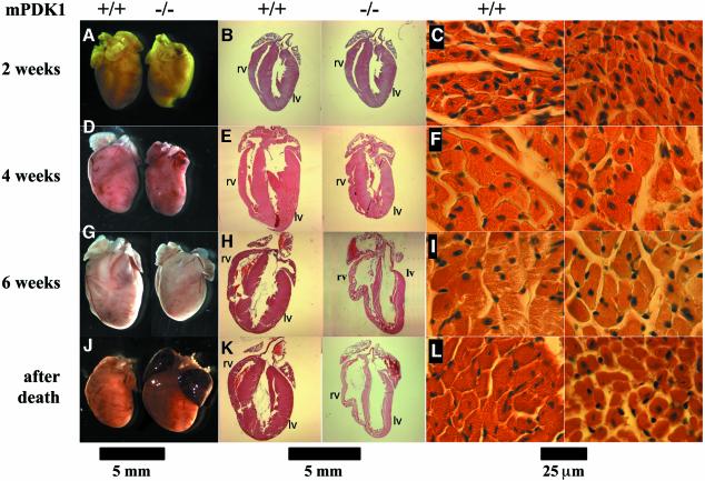
Fig. 2. Histological analysis of hearts from mPDK1+/+ and mPDK1+/+ mice. At the indicated times the hearts were fixed in 10% formalin, embedded in wax and stained with haematoxilin and eosin. (A, D, G and J) Comparison of a representative image of heart of littermate mPDK1–/– and mPDK1+/+ mice of the same sex, before the fixation and staining. (B, E, H and K) Representative longitudinal sections after fixation and staining. (C, F, I and L) Representative micrographs of transversal sections of the muscular fibres. Scale bar is shown at the bottom of each set of panels.

An official website of the United States government
Here's how you know
Official websites use .gov
A
.gov website belongs to an official
government organization in the United States.
Secure .gov websites use HTTPS
A lock (
) or https:// means you've safely
connected to the .gov website. Share sensitive
information only on official, secure websites.
