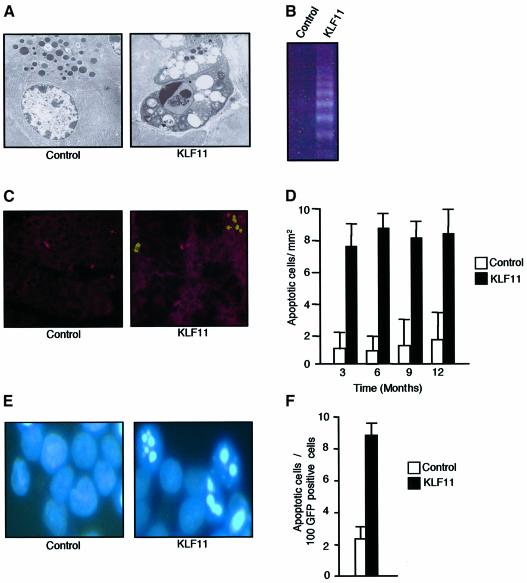
Fig. 4. KLF11 overexpression shows an increase in the rate of apoptosis both in vivo and in vitro. (A) Electron microscopic examination revealed typical signs of apoptosis in transgenic mice, such as the formation of cytoplasmic blebs, and apoptotic bodies were also found in transgenic mice in addition to chromatin condensation. (B) KLF11 transgenic mice showed the formation of a typical DNA ladder as a result of internucleosomal fragmentation. (C) TUNEL staining showing a representative pancreas section of 3-month-old KLF11 transgenic and wild-type mice. (D) To quantify the degree of apoptosis, we counted the number of TUNEL-positive cells per mm2 and calculated the change of apoptosis. As demonstrated, a 5-fold increase in the number of apoptotic cells was observed in KLF11 transgenic mice compared with wild-type mice. This increase in apoptosis was consistent in KLF11-overexpressing animals of all ages. (E) Nuclear Hoechst staining showed the typical morphological changes of the apoptosis KLF11-transfected PANC1 cells. (F) KLF11 overexpression induces 3- to 4-fold increases in the rate of apoptosis in PANC1 cells compared with cells transfected with the empty vector.

An official website of the United States government
Here's how you know
Official websites use .gov
A
.gov website belongs to an official
government organization in the United States.
Secure .gov websites use HTTPS
A lock (
) or https:// means you've safely
connected to the .gov website. Share sensitive
information only on official, secure websites.
