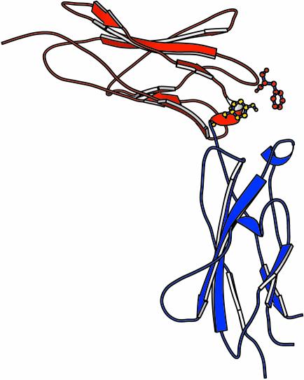
Fig. 4. Ribbon representation of the crystal structure of the gp130 CHD (Bravo et al., 1998). The locations of the two principal residues involved in ligand recognition via site II are presented in ball-and-stick format: Phe73 (red) and Tyr100 (blue). Residues are numbered according to the crystal structure.

An official website of the United States government
Here's how you know
Official websites use .gov
A
.gov website belongs to an official
government organization in the United States.
Secure .gov websites use HTTPS
A lock (
) or https:// means you've safely
connected to the .gov website. Share sensitive
information only on official, secure websites.
