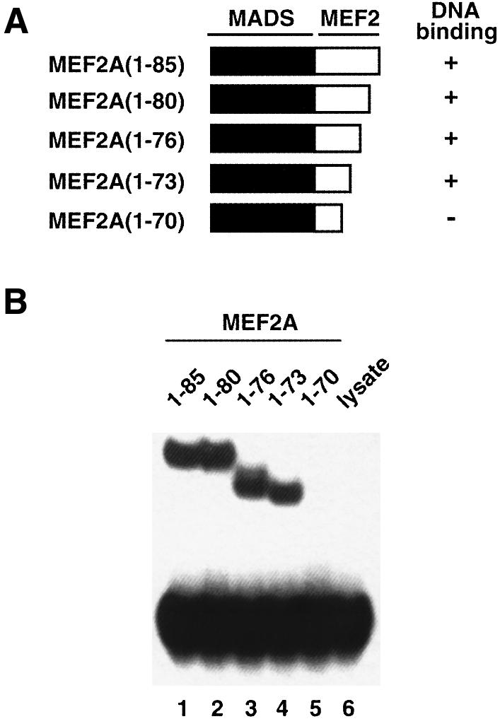
Fig. 4. Mapping the minimal MEF2A DNA-binding domain. (A) Schematic illustration of the truncated MEF2A constructs. (B) Gel retardation analysis of equimolar concentrations of in vitro translated MEF2A proteins bound to the N10 site (central motif CTATTTATAG; Sharrocks et al., 1993a). Approximately equal concentrations (∼50 nM) of protein and DNA were used. Lane 6 is a control containing unprogrammed rabbit reticulocyte lysate.
