
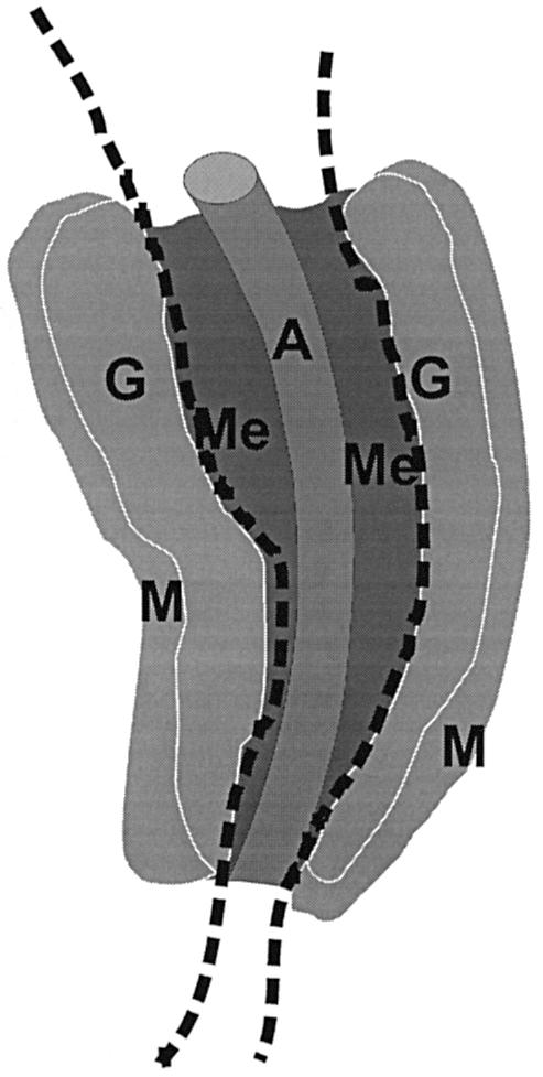
Fig. 1. Schematic representation of subdissected regions of the AGM. E11 and E12 AGM tissue is dissected along the dotted lines to separate the dorsal aorta and surrounding mesenchyme from the urogenital ridges, the latter of which contain the developing gonads and pro/mesonephroi. A, dorsal aorta; Me, mesenchyme; G, gonad; M, mesonephros.