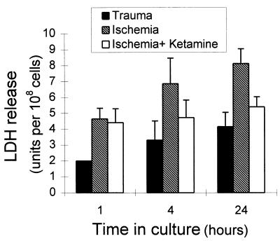
Figure 1.
LDH release by retinal tissue exposed to trauma or ischemia. Retinal tissue from E17 embryos was exposed to trauma by the process of excising the tissue and subsequently cutting it into pieces. The tissue was organ-cultured for 24 h, and LDH release was measured in samples of the culture medium 1, 4, and 24 h after tissue excision (solid bars). E17 retina was exposed to ischemia by organ-culturing the tissue pieces for 50 min in glucose-free medium in flasks that were gassed with 95% N2/5% CO2 (hatched bars). In some experiments the culture medium of the ischemic tissue contained the glutamate antagonist ketamine (10 mM/ml) (open bars). The levels of LDH were measured in medium samples 1, 4, and 24 h after tissue excision. Each bar represents the mean ± SD of three separate experiments, each performed in triplicate (n = 9).

