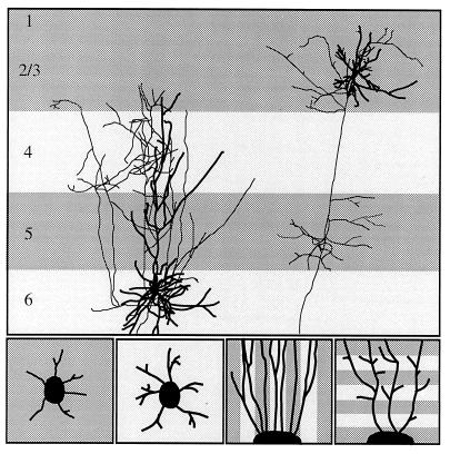
Figure 1.
(Upper) Schematic diagram of interlaminar connections of pyramidal cells in layers 2/3 and in layer 6. [The layer 6 cell is from M. Hübener and J.B., unpublished work; the layers 2/3 cell is reproduced with permission from ref. 31 (copyright 1994, by Cell Press).] Thick processes are dendrites, thin processes, axons. Notice that axon collaterals of the layer 6 cell target lightly shaded laminae (layer 6 and layer 4), whereas axon collaterals of the layers 2/3 cell arborize in the darkly shaded laminae (layers 2/3 and layer 5). (Lower) In vitro experiments designed to investigate the role of membrane-associated molecules on the formation of layer-specific cortical circuits. In the two panels on the left, explants of cortical cells destined either for layers 2/3 or for layer 6 are placed on a homogeneous membrane carpet from target and nontarget layers. In the two panels on the right, axons extending from cortical explants are confronted with alternating lanes of membranes prepared from target and nontarget layers. Axons grow either parallel to the membrane lanes (axonal guidance assay) or perpendicular to the lanes (axonal branching assay).

